Dermoscopy stands as a cornerstone in dermatological diagnostics, offering a window into the intricate world of skin lesions and abnormalities. This article delves into the nuances of dermoscopy, revealing how it magnifies the skin surface to detect and differentiate between benign and malignant conditions, with a particular focus on skin cancer. Through a detailed examination of the procedure and its clinical applications, we uncover the significance of dermoscopy in achieving early diagnosis and improving patient outcomes. Join us as we explore the essential role of dermoscopy in the early detection and management of skin cancer, enhancing the precision and efficacy of dermatological care.
Table of Contents
What is Dermoscopy?
Dermoscopy, also known as skin surface microscopy, is a diagnostic tool in dermatology for examining pigmented skin lesions and other skin conditions. It involves using a dermatoscope to magnify and illuminate the skin surface, providing a clear view of structures not visible to the naked eye. This technique is crucial for the accurate diagnosis of skin lesions, aiding in distinguishing benign growths from malignant melanoma and other types of skin cancer. The non-invasive nature of dermoscopy allows for quick and effective examination during routine check-ups, making it a fundamental practice in dermatology for early diagnosis.
What is Dermoscopy Used For?
Dermoscopy is instrumental in the early diagnosis and management of skin cancer, including malignant melanoma and basal cell carcinoma. It enables dermatologists to closely examine suspicious skin lesions and identify characteristics indicative of malignancy. Beyond skin cancer detection, dermoscopy is also used for diagnosing various dermatological conditions such as eczema, psoriasis, and fungal infections. The enhanced imaging of pigmented lesions through dermoscopy facilitates better clinical decisions and treatment planning.
What Are Dermatoscopes?
Dermatoscopes are advanced optical devices designed for skin surface microscopy. They magnify and illuminate skin lesions, allowing dermatologists to observe features like color patterns, vascular structures, and morphological details. Modern dermatoscopes often integrate digital technology, enabling the capture and storage of images for ongoing assessment and monitoring of pigmented skin lesions. This feature is particularly valuable for tracking changes in lesions over time, crucial for diagnosing primary melanoma and other skin conditions.
Can Dermoscopy Detect Cancer?
Dermoscopy is a key tool in detecting skin cancer. It can reveal detailed characteristics of skin lesions that are indicative of basal cell carcinoma, malignant melanoma, and other cancers.
By providing a magnified view of the lesion, dermoscopy enhances the dermatologist’s ability to detect cancerous changes at an early stage, significantly improving the chances of successful treatment. While highly effective, dermoscopy complements, rather than replaces, histopathological analysis for a definitive cancer diagnosis.
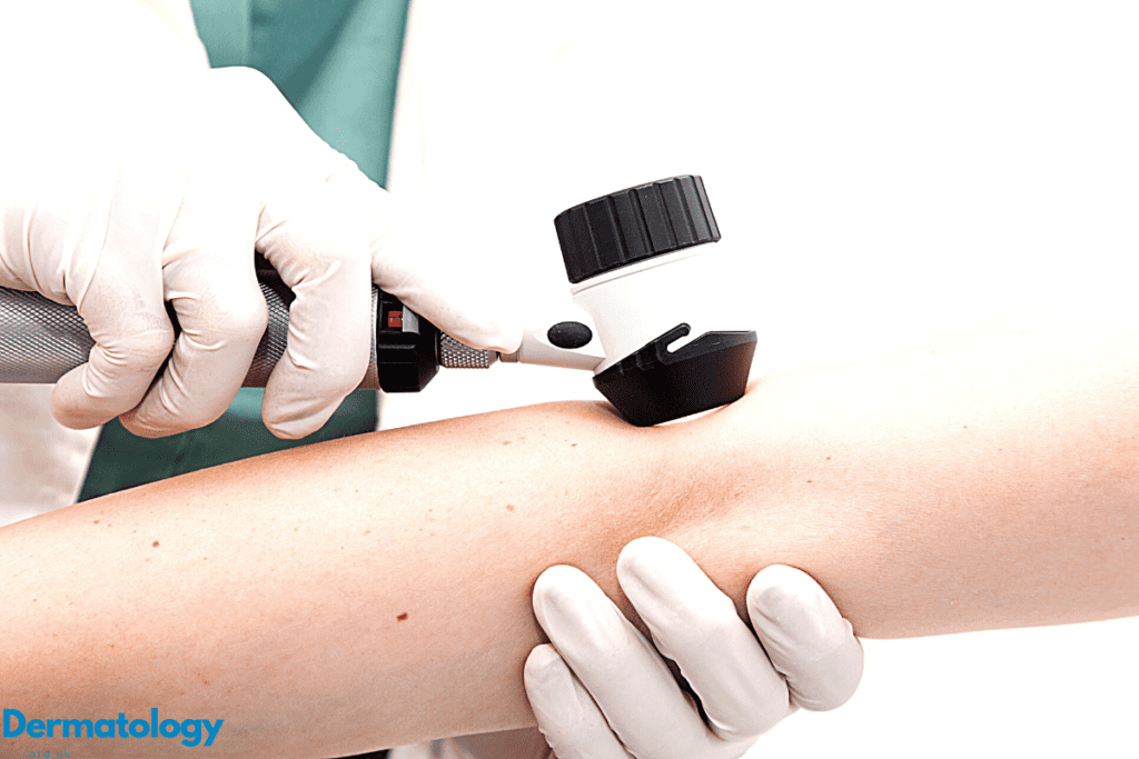
How is Dermoscopy Performed?
The procedure of dermoscopy involves placing a dermatoscope against the skin surface, often with a transparent medium like gel or oil to enhance optical clarity. Dermatologists then examine the skin lesions through the device, assessing characteristics such as symmetry, border, color, and pattern.
This detailed analysis assists in differentiating benign from malignant lesions, guiding the subsequent medical or surgical approach. The non-invasive and detailed nature of dermoscopy makes it an essential technique in dermatological examinations and skin cancer detection.
What Can You See with a Dermatoscope?
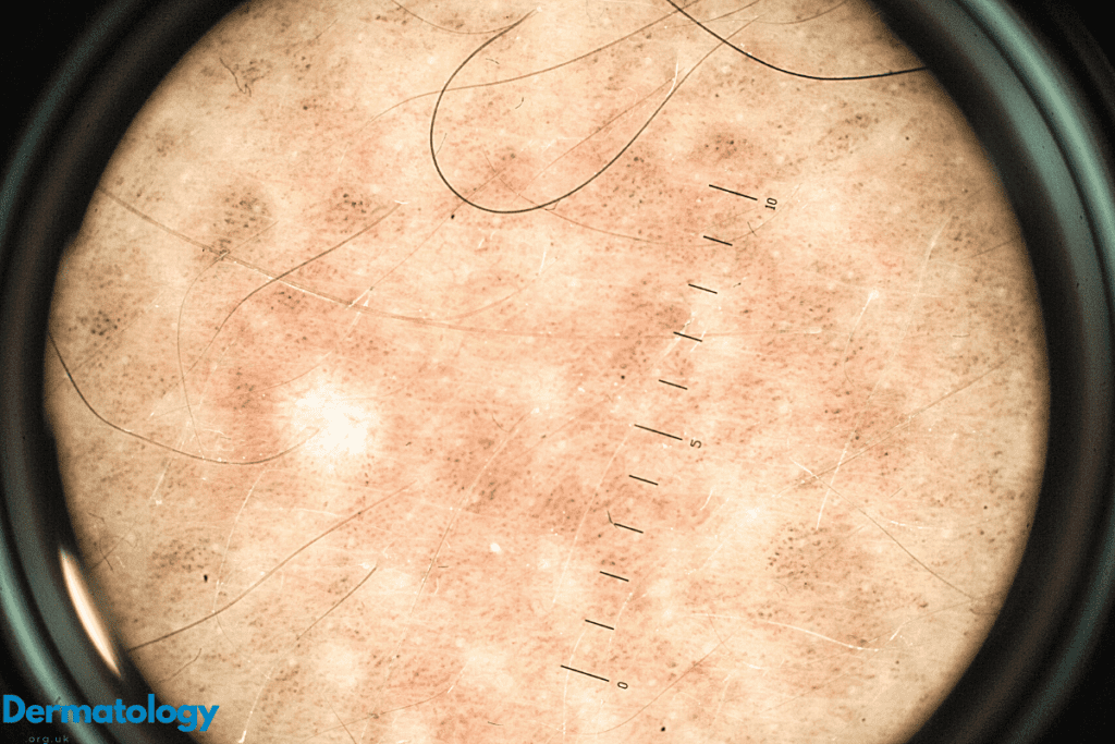
A dermatoscope provides a detailed view of the skin surface, revealing pigmented lesions, vascular patterns, and other skin anomalies. This close examination helps in diagnosing various skin conditions, from benign moles to malignant melanoma. Dermatoscopy exposes subtle details that are crucial for accurate diagnosis and effective treatment planning, making it an invaluable tool in the field of dermatology for the management of skin lesions.
How Long Does a Dermoscopy Take?
The duration of a dermoscopy examination can vary, typically taking a few minutes per lesion. However, the process may extend if multiple lesions require detailed assessment. Despite the potential for varying timeframes, dermoscopy remains a swift and efficient method for evaluating skin lesions, with the time invested being crucial for the early diagnosis and management of skin cancer.
Additional Insights into Dermoscopy
Enhancing Early Detection of Skin Cancer
Dermoscopy’s role extends beyond mere examination; it is pivotal in the early detection of skin cancer. Its ability to provide magnified, detailed images of skin lesions enables dermatologists to identify early signs of malignancy, particularly in pigmented skin lesions and cases of primary melanoma.
The Evolution of Dermatoscopic Technology
The advancement in dermatoscopic technology, including the integration of digital imaging and analysis, has significantly improved the diagnostic accuracy and efficiency of skin cancer detection. These technological enhancements allow for better tracking and monitoring of skin lesions, facilitating a proactive approach in the management of dermatological conditions.
Dermoscopy in Clinical Practice
Incorporating dermoscopy into routine clinical practice has revolutionized the approach to diagnosing and managing skin lesions. It provides a bridge between clinical examination and histopathological evaluation, offering a real-time, in-depth analysis of the skin’s surface. Dermoscopy has become an indispensable part of dermatological diagnostics, underscoring its importance in modern medicine.
Overview
Dermoscopy is a vital diagnostic technique in dermatology, enhancing the evaluation of skin lesions and aiding in the early diagnosis of skin cancer. With its ability to magnify and elucidate subtle skin details, dermoscopy serves as a critical tool in the early detection and management of skin cancer, particularly malignant melanoma and basal cell carcinoma. As technology advances, the role of dermoscopy in dermatological practice continues to expand, promising better outcomes for patients with skin conditions.